Microscope Smooth Muscle Diagram : 2 937 Muscle Cells Stock Photos Pictures Royalty Free Images Istock - Note that skeletal muscle cells are multinucleate, that is, each cell has more than one nucleus.
byAdmin-
0
Microscope Smooth Muscle Diagram : 2 937 Muscle Cells Stock Photos Pictures Royalty Free Images Istock - Note that skeletal muscle cells are multinucleate, that is, each cell has more than one nucleus.. Most of the muscle cell nuclei you see will be along the sides of the cells. Smooth muscle cells are spindle shaped, have a single, centrally. Slides of skeletal muscle come in a variety of forms. It is the pen diagram of skeletal, smooth and cardiac muscle for class 10, 11 and 12. Skeletal and cardiac muscle cells are called striated muscle because of the very regular arrangement of their intracellular contractile units, sarcomeres, at the light microscope (lm) and electron microscope (em) levels.
All slides are 40x or. Do not overlap (this is why these bands appear paler under the microscope). Smooth muscle (textus muscularis levis) smooth muscle is a type of tissue found in the walls of hollow organs, such as the intestines, uterus and stomach. About press copyright contact us creators advertise developers terms privacy policy & safety how youtube works test new features press copyright contact us creators. This pattern of light and dark is due to the arrangement of the protein filaments involved in muscle contraction.
Smooth Muscle Tissue Muscle Anatomy Smooth Muscle Tissue Human Skull Anatomy from i.pinimg.com This pattern of light and dark is due to the arrangement of the protein filaments involved in muscle contraction. Histology of human smooth muscle under microscope view for education. Smooth muscle can be confused easily with ordinary connective tissue. Note that skeletal muscle cells are multinucleate, that is, each cell has more than one nucleus. This diagram shows a few of the cells that can be seen in the stained section below. Note the striations (stripes) that run across each cell. Muscle tissues differ in structure. Smooth muscle is composed of sheets or strands of smooth muscle cells.
On the left is the view with light microscopy.
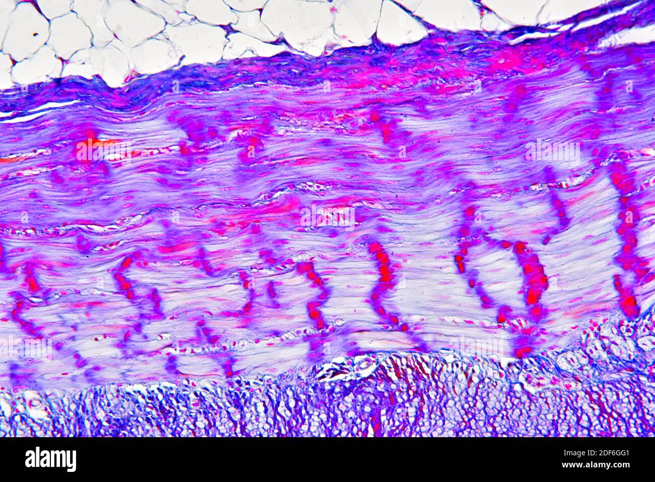
Smooth muscle can be confused easily with ordinary connective tissue. Diagram of contraction of skeletal muscle. Smooth muscle is specialized for slow and sustained contractions of low force. You can have a slide of pure skeletal muscle, or slides of various organs, such as tongue or larynx, that contain skeletal muscle. (a) observe slide of smooth muscle fibre under the microscope and draw its labelled diagram. Smooth muscle has inherent contractility, and the autonomic nervous system, hormones and local metabolites can influence its contraction. Muscle tissues differ in structure. In this video i have shown the simplest way of drawing muscle drawing. Smooth muscle and endothelium in a muscular artery wall, (magnification x100). The images show only part of length of these skeletal muscle cells. Skeletal muscle 400x the bar shows the width of one skeletal muscle cell. Smooth muscle has bundles of thin and thick filaments. The cells are spindle shaped, and the nucleus is central.
The images show only part of length of these skeletal muscle cells. Before placing your slide on the microscope. Smooth muscle has bundles of thin and thick filaments. It is the pen diagram of skeletal, smooth and cardiac muscle for class 10, 11 and 12. In this video i have shown the simplest way of drawing muscle drawing.
Anatomy Of A Skeletal Muscle Fiber Video Khan Academy from i.ytimg.com Since it is not under conscious. Muscular system tour lab page 4 the muscles … a front view. Smooth muscle is a type of muscle tissue which is used by various systems to apply pressure to vessels and organs. If you were to look at skeletal, smooth and cardiac muscle using a microscope, you would see differences in their structure. Skeletal muscle cells (also known as skeletal muscle fibers) are cylindrical, multinucleate and very long. Anatomy learner will provides more article like trachea histology slide in regular basis. Smooth muscle contracts under certain stimuli as atp is freed. Note the striations (stripes) that run across each cell.
These cells have fibers of actin and myosin which run through the cell and are supported by a framework of other proteins.
Histological features of animal lung with slide image and labeled diagram #2. This is the best guide to learn trachea histology with slide images and labeled diagram. (a) observe slide of smooth muscle fibre under the microscope and draw its labelled diagram. In one of his biceps muscle fibers at rest, the length of the i band is 1.0 μm and the a band is 1.5 μm. Smooth muscle diagram wiring diagram skeletal muscle definition function britannicacom. In this video i have shown the simplest way of drawing muscle drawing. When you see the skeletal muscle in the microscope you will see that there is a different structure. This pattern of light and dark is due to the arrangement of the protein filaments involved in muscle contraction. It constitutes much of the musculature of It is the pen diagram of skeletal, smooth and cardiac muscle for class 10, 11 and 12. Use your front view and back view This diagram shows a few of the cells that can be seen in the stained section below. The muscles … a back view.
(muscle cells are often referred to as muscle fibers because of their narrowness and length.). If you were to look at skeletal, smooth and cardiac muscle using a microscope, you would see differences in their structure. Under the microscope note that fascicles of smooth muscle are arranged in various planes. It is the pen diagram of skeletal, smooth and cardiac muscle for class 10, 11 and 12. This diagram shows a few of the cells that can be seen in the stained section below.
Smooth Muscle High Resolution Stock Photography And Images Alamy from c8.alamy.com Histology of human smooth muscle under microscope view for. This is the best guide to learn trachea histology with slide images and labeled diagram. Do not overlap (this is why these bands appear paler under the microscope). Muscle is one of the four basic types of tissue and is specialized for contraction. Histology of human smooth muscle under microscope view for education. Smooth muscle and endothelium in a muscular artery wall, (magnification x100). Smooth muscle is the simplest of the three kinds of muscle. It is the pen diagram of skeletal, smooth and cardiac muscle for class 10, 11 and 12.
Anatomy learner will provides more article like trachea histology slide in regular basis.
There are three major types of muscle, and their structure reflects their function. Skeletal muscle fibres are packed into regular parallel bundles. The nucleus identified in the image (nuc) is just inside the cell membrane, but the top the cell was caught by chance in this section. Muscle is one of the four basic types of tissue and is specialized for contraction. When you see the skeletal muscle in the microscope you will see that there is a different structure. The images show only part of length of these skeletal muscle cells. (b) give name of atleast two structures where smooth muscle fibres are present in humans. Slides of skeletal muscle come in a variety of forms. Smooth muscle contracts under certain stimuli as atp is freed. Smooth muscle can be confused easily with ordinary connective tissue. Look at this slide grossly and at the microscope level. Since it is not under conscious. Smooth muscle cells are spindle shaped, have a single, centrally.